
Team Giannone in Nature Cell Biology, highlighted in Science
A study by the Team Spatio-temporal and mechanical control of motile structures (IINS) in collaboration with Institut Curie.
Cells sense mechanical forces and convert them into biochemical signals through dedicated molecular architectures. Among them, caveolae are small bulb-shaped invaginations of the plasma membrane. Long studied for their roles in signalling and trafficking, these multifunctional nanodomains are also key membrane mechanosensors: by flattening out, caveolae buffer sudden increases in membrane tension and protect the plasma membrane when cells are mechanically stressed by fluid flow (endothelial cells) or contraction/elongation (muscle cells). Whether mechanical forces acting on caveolae are translated into regulation of intracellular signals, however, remained unknown.
In a recent article published in Nature Cell Biology and highlighted in Science, Grégory Giannone, Olivier Rossier and several team members (IINS) joined their forces with Christophe Lamaze and Cédric Blouin (Institut Curie, Paris) for a collaborative study to investigate how the mechanics of caveolae regulate signalling pathways.
Combining single-molecule imaging and super-resolution microscopy, they found that mechanical stress rapidly disassembles caveolae and releases caveolin-1 scaffolds, oligomeric assemblies of the main caveolar protein, which then diffuse freely across the plasma membrane. Diffusing caveolin-1 scaffolds with exposed scaffolding domains can bind directly to the tyrosine kinase JAK1, inhibiting its catalytic activity. The same tension-controlled mechanism applies to at least three other signalling enzymes: eNOS, PTEN and PTP1B. A theoretical model based on the thermodynamics of caveolae assembly reproduced these observations, supporting the generality of the mechanism.
Together, this study defines a new mechanotransduction paradigm in which signalling information is decoded remotely from the initial mechanosensing caveola, as tension-released caveolin-1 scaffolds diffuse away and form dynamic reversible complexes with signalling effectors elsewhere on the membrane.
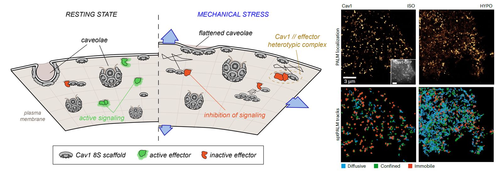
Figure:
Left: graphical abstract: Upon mechanical stress (cell stretching or osmotic shock), caveolin-1 scaffolds are released from caveolae and undergoes lateral diffusion along the plasma membrane where they form dynamic reversible complexes with signalling effectors (green, active signaling; red, inhibition of signaling).
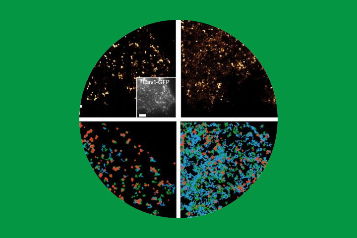
Right: Top shows representative super-resolution PALM images of caveolin-1 (Cav1) in a mouse lung endothelial cell under isotonic conditions (left, ISO) and during hypotonic shock (right, HYPO). Inset shows corresponding diffraction-limited images of coexpressed Cav1–GFP. Bottom shows colour-coded single-particle trajectories of Cav1 classified as diffusive (blue), confined (green) or immobile (red) in the same cell before (left) and during hypotonic shock (right).
For more information
Diffusing caveolin-1 scaffolds regulate mechanosignalling, Satish Kailasam Mani†, Nicolas Tardif†, Olivier Rossier†, Ismail M. Khater, Xuesi Zhou, Victor Breton, Filipe Nunes Vicente, Adiyodi Veettil Radhakrishnan, Céline Gracia, Pamela Gonzalez Troncoso, Isabel Brito, Richard Ruez, Melissa Dewulf, Ghassan Hamarneh, Ivan Robert Nabi, Philippe Cuniasse, Pierre Sens, Grégory Giannone‡, Cédric M. Blouin‡* & Christophe Lamaze‡*
† These authors contributed equally to this work (co-first authors).
‡ These authors jointly supervised this work (co-senior authors).
* Corresponding authors
Nature Cell Biology, June 1st 2026. DOI: 10.1038/s41556-026-01966-0
Highlighted in Science, DOI: 10.1126/science.aej7459
Contact
Grégory Giannone
CNRS Researcher – Team leader
Team Spatio-temporal and mechanical control of motile structures
Olivier Rossier
INSERM Researcher
Team Spatio-temporal and mechanical control of motile structures
Last update 01/07/26