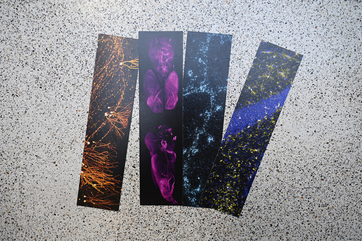
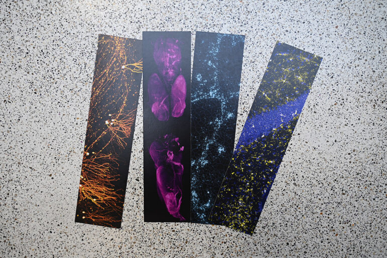
A new addition to our bookmark collection, this time featuring an image by Céline Lucas (NutriBrain) and Jean-Christophe Delpech (NutriNeuro – Université de Bordeaux, INRAE, Bordeaux INP).
Don’t hesitate to pick some up at the Hippocampus office!
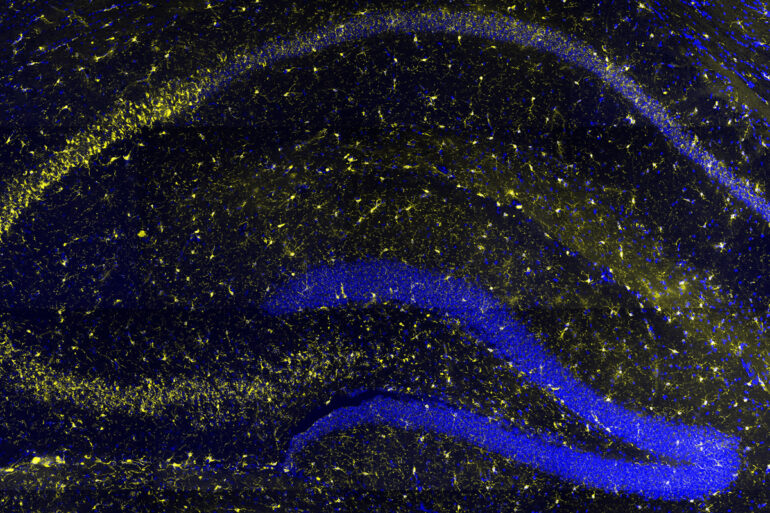
Pictures used :
Picture: Granular neurons of the dentate gyrus generated in an adult mouse and labelled thanks to an injection of retrovirus coding for GFP and after clearing of the brain. Credits: Pierre Mortessagne, Emilie Pacary (Neurocentre Magendie, Univ. Bordeaux, INSERM) and Jérémie Teillon (Bordeaux Imaging Center, Univ. Bordeaux, CNRS, INSERM)
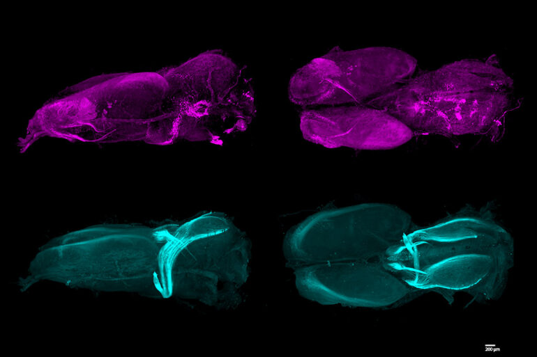
Picture: Three-dimensional visulation of catecholaminergic innervation in an entire adult newt’s brain (Pleurodel Waltl). Credits: Aude Retailleau (IMN – Univ. Bordeaux, CNRS) in collaboration with Anaïs Favre and Alain Chedotal (Institut de la Vision – Sorbonne Université, INSERM, CNRS).
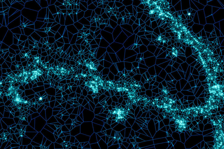
Picture: AMPA receptor labeling in primary hippocampal neurons. Credits: Hanna Zieger (IINS – Univ. Bordeaux, CNRS). Software used: SR-Tesseler developed by Florian Levet (BIC – Univ. Bordeaux, CNRS, Inserm / IINS – Univ. Bordeaux, CNRS)
Picture: Microglial cells in hippocampus of aged mice (Iba1 and DAPI staining, Microscope Axio Observer 7, appotome epifluorescent, x20). Credits: Céline Lucas (NutriBrain) and Jean-Christophe Delpech (NutriNeuro – Univ. Bordeaux, INRAE, Bordeaux INP).