
Endocytosis in the axon initial segment maintains neuronal polarity
In a study published in Nature, researchers from the “membrane traffic at synapses” team, led by Dr David Perrais at the Institute of Interdisciplinary Neuroscience (CNRS and University of Bordeaux), collaborated with teams from Stanford University in California to identify a third mechanism for preserving neuron polarity: the internalization of dendritic surface proteins by endocytosis and their degradation in the AIS. To reach this conclusion, the researchers used three biological models. Firstly, in the Stanford team led by Professor Kang Shen, Dr Kelsie Eichel studied certain neurons in the nematode worm Caenorhabditis elegans, a one-millimeter long worm that is one of the most studied model systems by biologists. Focusing their study on one of the 302 neurons of this worm, they showed that this neuron possesses an AIS and that blocking endocytosis specifically in this neuron causes the redistribution of a dendritic surface protein called DMA-1 towards the axon. Furthermore, this protein, which is responsible for the formation of dendritic branches, causes ectopic formation of branches normally absent in the axon when it is present in the axon. Eichel and colleagues have also shown that endocytosis plays the same role for dendritic surface proteins in human neurons, and also in rodent neurons based on experiments done in collaboration with Professors Marius Wernig, Robert Malenka, and Nobel laureate Thomas Südhof and their teams at Stanford. The Bordeaux team made key contributions to this work by direct demonstration of AIS endocytosis using a fluorescence imaging method to detect endocytosis vesicle formation in real time (see Sposini et al. 2020) to show that dendritic surface proteins are indeed internalized in the AIS of neurons, and that the frequency of endocytosis vesicle formation is the same as in dendrites.
These results constitute an important advance in the understanding of the cellular mechanisms of neuronal polarity formation and preservation. They will also allow us to study this new form of endocytosis and to determine its specificity mechanisms, which could lead to a better understanding of the functioning of the AIS, an essential compartment of neuronal function.
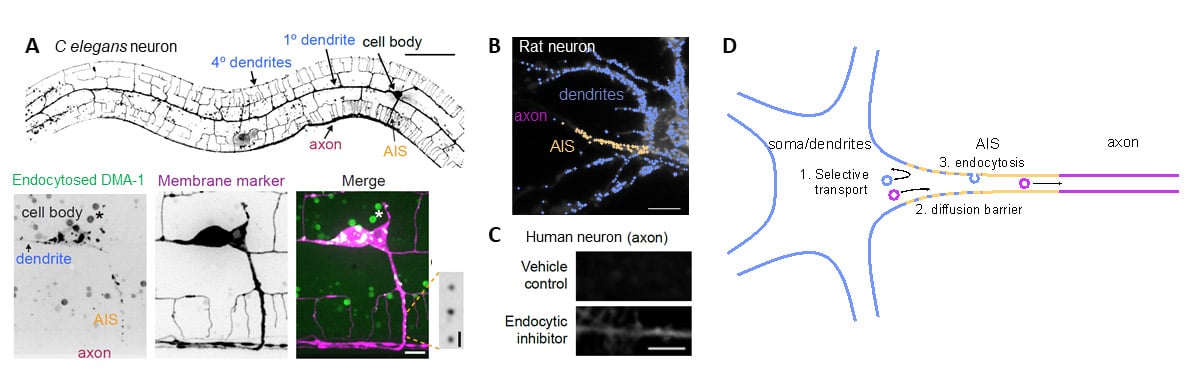
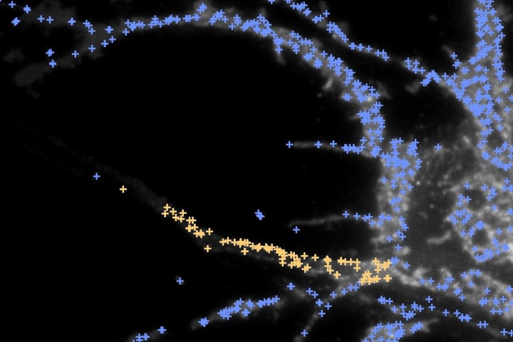
Figure: Endocytosis at the AIS is necessary for maintaining the polarity of dendritic proteins. A, Morphology of the PVD neuron in C elegans. Dendrites are very long and branched, while the axon does not branch. In the AIS, identified with various markers, dots mark endocytosed dendritic protein (GFP-FLPon-CLIC1). B, In a rat neuron, endocytic events containing a dendritic protein occur in the dendrites and soma (light blue crosses) and the AIS (yellow crosses) but very few in the axon. C, In human neurons, after blocking endocytosis dendritic proteins, normally not present in the axon, become detectable in the axon. D, Scheme of the three cellular mechanisms which maintain polarity in neurons. The third mechanism, endocytosis in the AIS, is revealed in the study by Eichel et al.
Articles
Endocytosis in the axon initial segment is a clearance mechanism to maintain neuronal polarity.
Kelsie Eichel, Takeshi Uenaka, Vivek Belapurkar, Rui Lu, Shouqiang Cheng, Joseph S. Pak, Caitlin A. Taylor, Thomas C. Südhof, Robert Malenka, Marius Wernig, Engin Özkan, David Perrais & Kang Shen
Nature ; 17 august 2022.
DOI : 10.1038/s41586-022-05074-5
Imaging endocytic vesicle formation at high spatial and temporal resolutions with the pulsed-pH protocol.
Silvia Sposini, Morgane Rosendale, Léa Claverie, Thi Nhu Van, Damien Jullié, David Perrais
Nature Protocols ; September 2020
DOI : 10.1038/s41596-020-0371-z
Contact
David Perrais
Chercheur CNRS à l’Institut interdisciplinaire de neurosciences (CNRS/Université de Bordeaux)
+33 5 33 51 48 61
Last update 07/09/22