
Gabriel Pitollat and al in AJRCCM
Congenital Central Hypoventilation Syndrome (CCHS) is characterized by a loss of ventilatory chemosensitivity and life-threatening hypoventilation. Patients require lifelong mechanical ventilation, at least nocturnally. Central apneas are commonly reported in CCHS patients, whereas obstructive apneas may remain unnoticed due to tracheostomy and positive pressure ventilation. CCHS is caused by mutations in the paired-like homeobox 2B gene (PHOX2B), the master gene of the autonomic nervous system. In mice, introduction of the most prevalent mutation observed in human (7-alanine expansion, PHOX2B27Ala/+) is associated with the main symptoms of CCHS, and agenesis of the retrotrapezoid nucleus, known to provide a major excitatory drive to breathing.
In this publication in AJRCCM we present our work, performed in collaboration with Dr. Jorge Gallego and Boris Matrot (Inserm U1141, Hôpital Robert Debré, Université de Paris, Paris) providing new insights in breathing disorders associated with CCHS. Combining pneumotachography and laser detection of abdominal movements (Figure 1A, B), to classify apneas, we demonstrate that newborn Phox2b27Ala/+ mice display an abnormally high degree not only of central apneas, but also of obstructive and mixed apneas (Figure 1C, D). Furthermore, these mice show dysgenesis (Figure 2) and dysfunction of the hypoglossal nucleus, a non-Phox2b expressing structure critical to the maintenance of upper airway patency. These results broaden the spectrum of neuroanatomical and neurofunctional disorders caused by Phox2b mutations, and point to the clinical importance of also detecting obstructive events in CCHS patients.
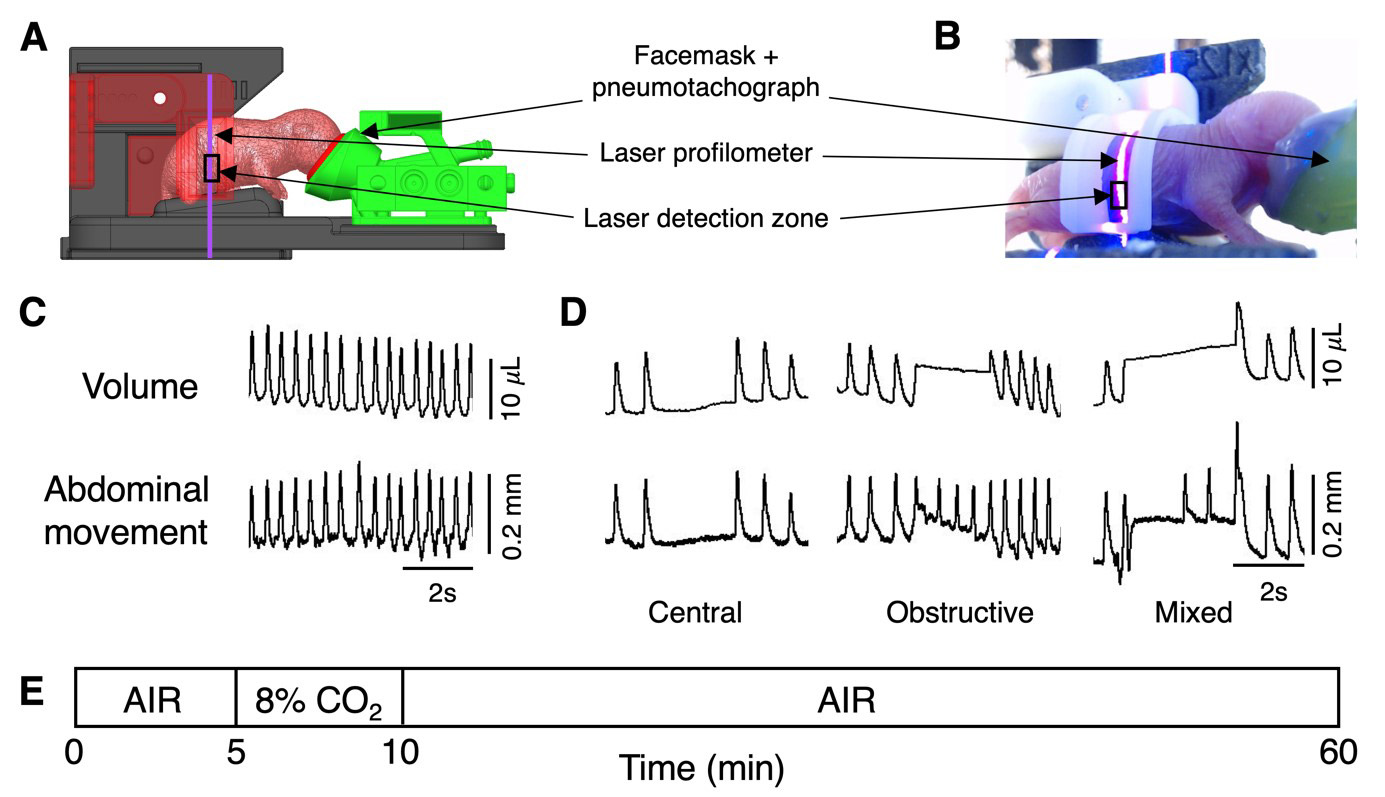
Figure 1: Different types of apneas are detected in CCHS pups. A-B: Experimental set up. C-D: Traces of flow and simultaneous abdominal movements during the different types of apneic events: central: no respiratory movement, no air flow; obstructive: respiratory movement, no air flow; mixed: some respiratory movements associated or not with air flow.
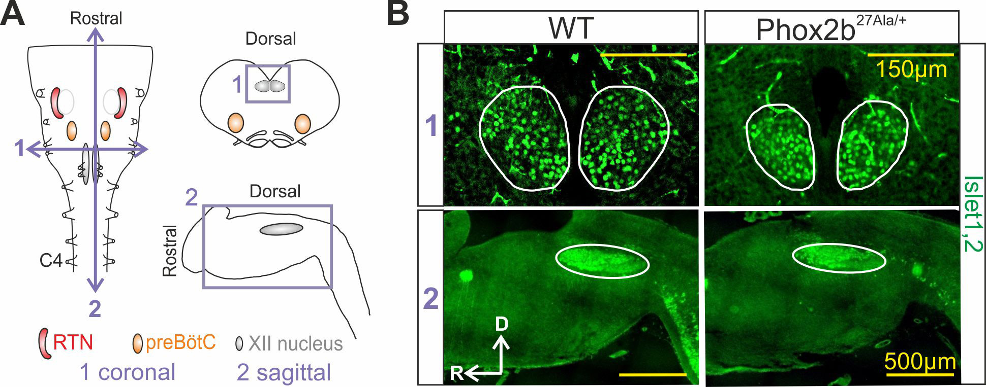
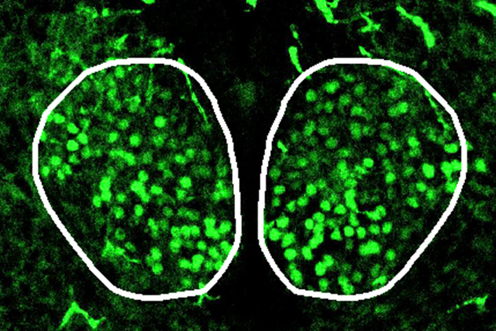
Figure 2: Abnormal anatomy of the hypoglossal nucleus in CCHS mutants. A: Schematic representation of brainstem preparations with the location of the main respiratory neural networks (RTN: retrotrapezoïd nucleus; preBötC: preBötzinger complex). B: Immunostaining against motoneurons (Islet1,2 positive) at the level of the hypoglossal nucleus in wild-type and CCHS mutant (Phox2b27Ala) preparations showing a reduced hypoglossal nucleus.
Reference
Obstructive Apneas in a Mouse Model of Congenital Central Hypoventilation Syndrome
Amélia Madani*, Gabriel Pitollat*, Eléonore Sizun, Laura Cardoit, Maud Ringot, Thomas Bourgeois, Nélina Ramanantsoa, Christophe Delclaux, Stéphane Dauger, Marie-Pia d’Ortho, Muriel Thoby-Brisson, Jorge Gallego, Boris Matrot.
Am J Respir Crit Care Med . 2021
doi: 10.1164/rccm.202104-0887OC.
* First co-authors
First co-author
Gabriel Pitollat (PhD student at INCIA) is funded with a Cifre grant in association with AtmosR
Mise à jour: 22/11/21