D. Choquet, M. Sainlos and JB. Sibarita in Nature Reviews Neuroscience
Abstract
The brain is arguably the most complex organ. The branched and extended morphology of nerve cells, their subcellular complexity, the multiplicity of brain cell types as well as their intricate connectivity and the scattering properties of brain tissue present formidable challenges to the understanding of brain function. Neuroscientists have often been at the forefront of technological and methodological developments to overcome these hurdles to visualize, quantify and modify cell and network properties. Over the last few decades, the development of advanced imaging methods has revolutionized our approach to explore the brain. Super-resolution microscopy and tissue imaging approaches have recently exploded. These instrumentation-based innovations have occurred in parallel with the development of new molecular approaches to label protein targets, to evolve new biosensors and to target them to appropriate cell types or subcellular compartments. We review the latest developments for labelling and functionalizing proteins with small localization and functionalized reporters. We present how these molecular tools are combined with the development of a wide variety of imaging methods that break either the diffraction barrier or the tissue penetration depth limits. We put these developments in perspective to emphasize how they will enable step changes in our understanding of the brain.
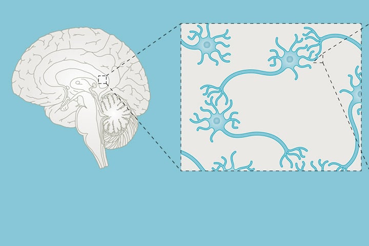
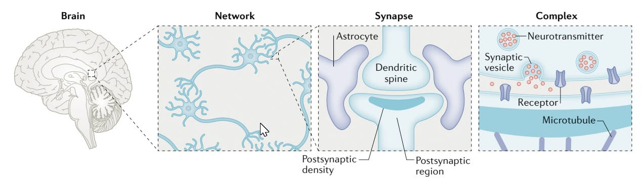
The different levels of complexity of the structures composing the brain are shown from the whole brain down to the molecular level, going through the neural network and synaptic levels.
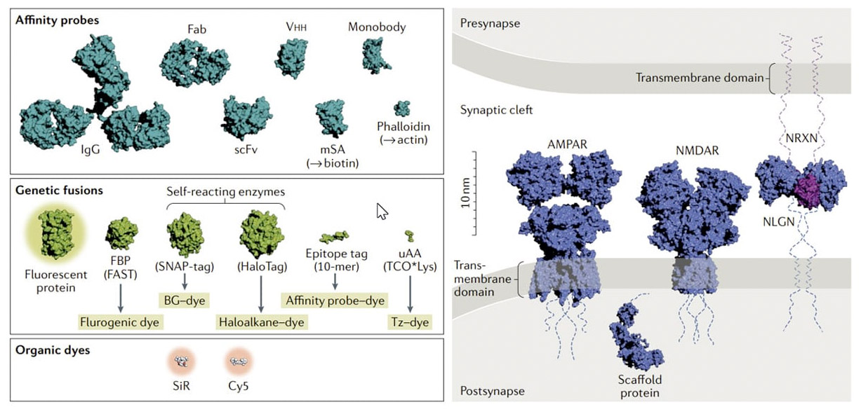
Left: Molecular structures of common affinity probes, tags (genetic fusions), organic dyes and representative neuronal targets all represented on the same scale. To image affinity probes, these are generally further functionalized with particles (not shown, from ~5 nm to a few tens of nanometres), fluorescent proteins or organic dyes. Affinity probes can either be developed against any protein target or be specific for a molecular target (specified in parentheses). Genetic fusion tags either are intrinsically fluorescent (fluorescent proteins) or require the addition of a fluorescent or fluorogenic module. Right: Illustrative neuronal targets consist of glutamate receptors, adhesion protein complex and scaffold proteins (dashed lines represent the non-resolved part of the complexes) represented in the confined environment of the synaptic cleft (~20–25 nm). The respective sizes of the various probes and targets in their biological context provide insights into the impact of the labelling strategy on parameters such as target accessibility, labelling density, distance between the fluorophore and the target (or linkage error) and impact on target function. Structures were obtained from the Protein Data Bank (IgG antibody, 1IGT; monovalent antibody fragment (Fab), 6NJM; single-chain variable fragment (scFv), 6J9O; single-domain antibody or nanobody (Vhh), 3G9A; monomeric streptavidin (mSA), 4JNJ; monobody, 3K2M; phalloidin, 6T25; fluorescent protein (GFP), 3G9A; fluorogen-based probe (FBP) FAST, 1NWZ; SNAP-Tag, 3KZY; HaloTag, 5UY1; epitope tag, 5IVN; trans-cyclooct-2-ene-derived lysine (TCO*Lys), 6AAO; AMPA receptor (AMPAR) complex, 6NJM; NMDA receptor (NMDAR), 6MMA; neuroligin (NLGN)–neurexin (NRXN) complex, 3BIW; scaffold protein (PSD95), derived from 2XKX, 3GSL, 3JXT, and 1JXO). BG, benzylguanine; 10-mer, 10-base oligonucleotide; Tz, tetrazine; uAA, unnatural amino acid.
Reference
Left to right : M. Sainlos, D. Choquet, JB Sibarita